
【探秘】前列腺应用解剖精粹(二)
编者按
在过去的几十年中,随着对前列腺解剖认识的不断加深以及手术技术的飞速发展,前列腺癌手术治疗的肿瘤控制和功能结果得到了显著改善。为帮助泌尿科医生了解根治性前列腺切除术涉及的解剖结构,并正确应用其中的命名术语,European Urology杂志发文(PMID:19931974)对前列腺及其周围结构作了详细描述。
本专栏对原文作了编译,并对原文图片作了高清化和中文标注。全部内容分为3期,本文为第二期,介绍前列腺周围筋膜(前列腺后方筋膜即Denonvilliers筋膜)、前列腺筋膜与神经血管束的关系、前列腺包膜、神经血管束(NVB)。因译者水平有限,译文难免存在不准确的地方,欢迎各位同道指正。
01 Posterior prostatic fascia and seminal vesicles fascia (Denonvilliers’ fascia)(前列腺后方筋膜和精囊筋膜(狄氏筋膜))
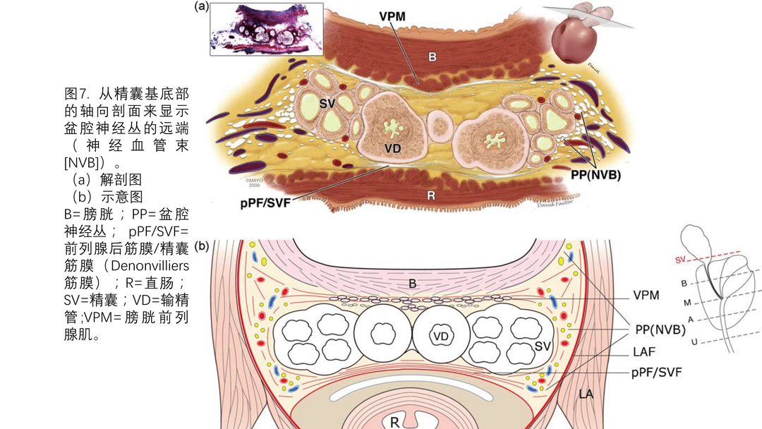
前列腺和精囊的后表面被一层连续的前列腺后方筋膜(pPF)和精囊筋膜(SVF)紧密覆盖,也被称为直肠前列腺筋膜、直肠膀胱隔、前列腺精囊筋膜和众所周知的狄氏筋膜(图1,2和7)。一些作者认为pPF/SVF起源于膀胱后穹隆处胚胎腹膜的融合。另有人认为pPF/SVF实际位于两层腹膜融合后残余物的前方。两层腹膜融合后使得后方的直肠固有筋膜与前方的pPF/SVF之间形成直肠前分离平面。
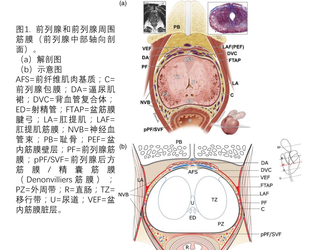
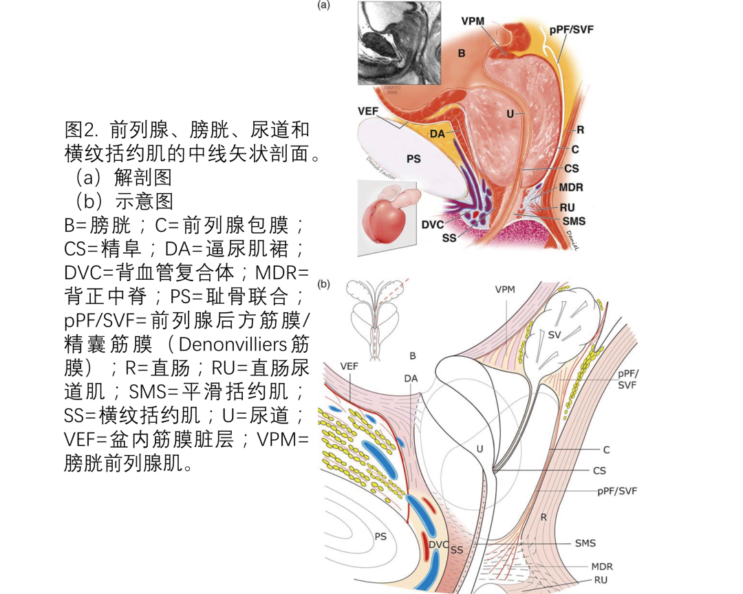
pPF/SVF头侧起源于腹腔尾端(直肠膀胱陷凹,图2)的前方。然后pPF/SVF向远端延伸至前列腺尖部,终止于前列腺尿道交界处,形成一个与会阴中心腱连续的终板。它由胶原纤维、弹性纤维和大量肌纤维构成,有着从易碎的半透明膜到致密的单层膜的变化。常提到的pPF/SVF的后层实际是直肠固有筋膜,它是独立于pPF/SVF的结构。pPF/SVF通常在前列腺的后表面中央与前列腺包膜融合。在前列腺的后外侧,pPF/SVF与前列腺包膜没有明显附着,在pPF/SVF与包膜之间的间隙内填充着网状组织和NVB。
02 Relationship between the prostatic fascias and the neurovascular bundle(前列腺筋膜与神经血管束的关系)
PF与NVB之间的关系存在争议。一些作者认为NVB完全位于前列腺包膜与LAF或pPF/SVF之间。在LAF的外侧或pPF/SVF的背侧未发现神经纤维。Kourambas等人对这种直截了当的观点提出了质疑,并指出在轴面上,pPF/SVF实际是前列腺两侧H形筋膜结构的一部分。左侧和右侧的PPF代表“H”的上肢,直肠旁筋膜代表“H“的下肢。“H”的横线对应的是pPF/SVF。他们的主要观察结果是pPF/SVF的外侧边界无明确界线,即三条筋膜相交处。他们观察到pPF/SVF横向分为数层,并在其中发现了神经纤维。这些神经纤维不仅位于pPF/SVF的腹侧,还位于pPF/SVF的背侧,而早先报道是无法辨认出这些神经纤维。这些没有明确界线的神经纤维层支持了NVB存在分隔和可能的功能分区。该发现也得到其他人证实,他们认为pPF/SVF实际是汇入NVB,或者从实际角度来看,pPF/SVF外侧缘分裂为前叶和后叶围绕NVB,并与LAF形成一个束缚NVB的三角形(图1)。
03 Prostate capsule(前列腺包膜)
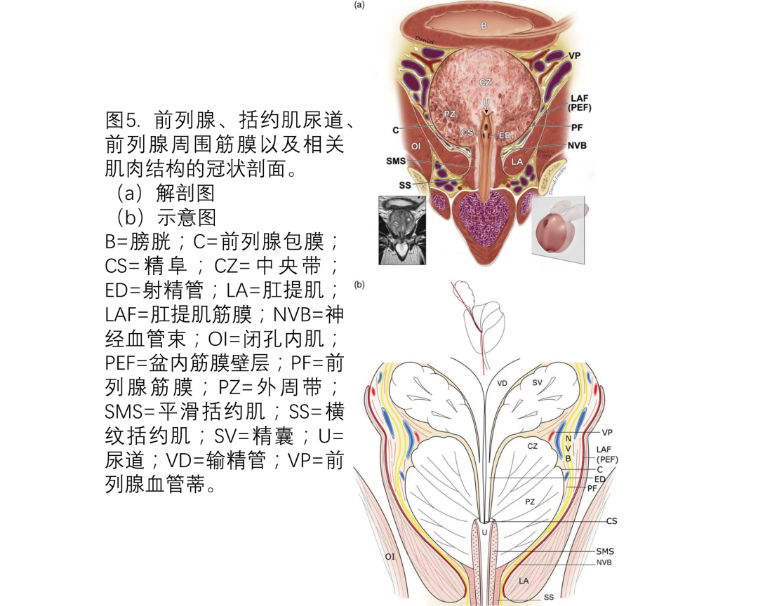
前列腺本身被一个囊状结构围绕,代表其外部边界。这个结构并不是解剖学意义上界线明确的囊,而是一层纤维肌束,主要是平滑肌,是前列腺基质不可分割的组成部分(图1和5)。多条血管和神经从前列腺外侧穿透前列腺包膜。通常不能在前列腺前面辨认出前列腺包膜,因为那里有前纤维肌肉基质和逼尿肌群。此外,也不能在前列腺尖部和基底部辨认出前列腺包膜。前列腺基质在前列腺尖部与尿道括约肌的肌纤维混合,在前列腺基底部与膀胱逼尿肌的平滑肌纤维混合。值得注意的是,尽管在前列腺解剖领域有着广泛研究,但围绕前列腺的筋膜的确切解剖仍存在争议。如前所述,我们建议将PPF简化为外层的LAF和内层的PF,后者覆盖前列腺包膜,神经血管结构并入或夹在LAF与PF之间,并且NVB存在个体差异。
04 Neurovascular bundle(神经血管束)
在男性,下腹下神经丛或盆腔神经丛负责勃起、射精和尿控机制。负责射精的交感神经纤维主要来自起源于T11–L2神经节的下腹神经。盆腔神经丛还含有副交感神经纤维(包括勃起神经),主要来自起源于S2-4腹支的盆腔和骶内脏神经。在海绵体中,这些神经主要负责勃起时血管扩张和动脉血流增加。盆腔神经丛的密集神经网络位于膀胱与直肠之间呈矢状走行的扁平状矩形纤维-脂肪层内。泌尿生殖道的神经分支走行在膀胱和膀胱颈的外侧并向尾侧延伸。盆腔神经丛的前部发出膀胱、前列腺、精囊和输精管的神经纤维,下半部发出负责勃起功能的海绵体神经。
盆腔神经丛的纤维以笼状围绕膀胱颈、前列腺近端和精囊的外侧,而在这些器官的前表面,神经纤维相对较少(图2,5,7,8)。这些神经纤维有些是微观的,在手术中无法被识别。然而在根治性前列腺切除术中,具有光学放大作用的手术技术能够更好地识别这些结构。尽管这些神经的确切作用尚不清楚,但很可能是直接支配这些器官。Costello等指出,这些走行至尿道括约肌、被命名为“海绵体神经”的分支主要位于盆腔神经丛的后部。它们位于精囊的后外侧,靠近精囊顶端走行。在根治性前列腺切除术中轻柔地游离精囊或采用保留精囊的技术可以减少损伤这些神经的风险,以改善术后尿控和勃起。据报道,这些神经与下列解剖标志的距离为:与精囊直接接触(0mm),距膀胱颈4mm,距肛提肌2mm。从这开始,海绵体神经和盆腔神经丛继续向尾侧走行,距前列腺血管蒂外侧仅0-7mm。这些神经纤维往往保持微观,并伴有血管结构,这就是为什么将该复合体称为NVB。该神经束不仅包含支配阴茎海绵体的神经纤维,还包含支配前列腺和尿道括约肌的神经纤维。从前列腺基底部到尖部,NVB的血管分出终末支穿透前列腺包膜,将NVB与包膜固定。
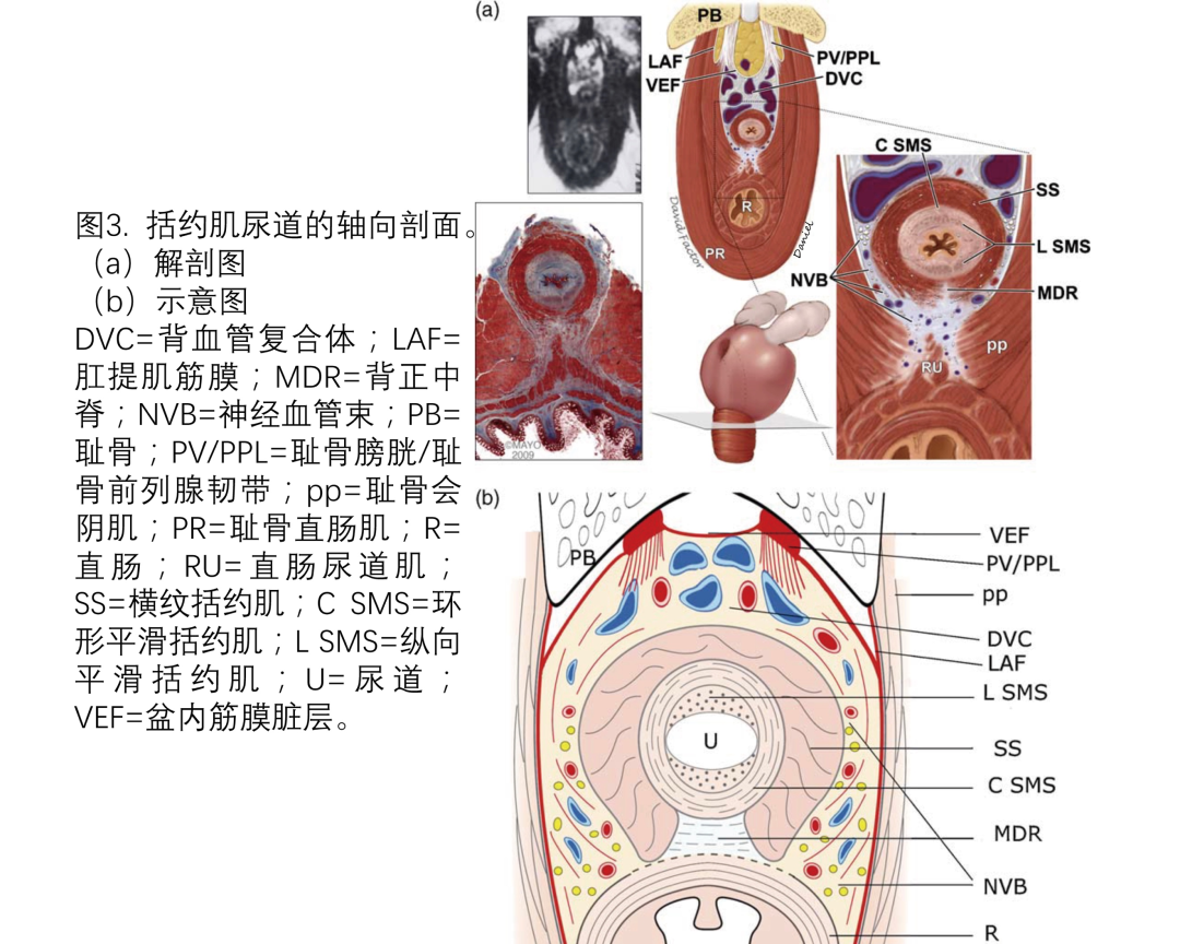
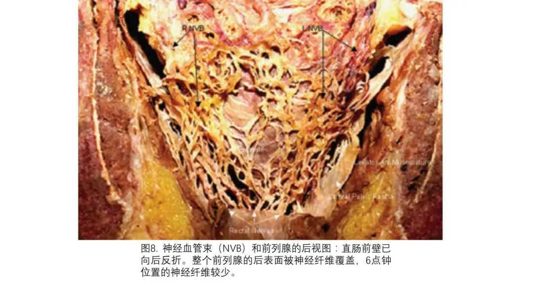
前列腺外侧的神经解剖可能会有很大变化。Müller的最初报告描述了位于前列腺后外侧和前外侧的神经。Walsh和Donker随后描述了位于前列腺后外侧的明显的NVB。最近的研究表明,该区域的神经解剖存在差异。Takenaka等证实了如Muller最初描述的那样,神经在前列腺的外侧和前外侧表面呈喷雾状分布,并得出结论,NVB不是一个明显的结构,而是由许多细微分散的纤维组成。Lunacek等发现,NVB在妊娠胚胎期发育为明显的结构,随后在前列腺发育时分散在前列腺外表面。他们还证实了前列腺增生的进展大大增加了神经在前列腺外表面的分散。然而Ganzer等发现,前列腺外表面的神经数量不受前列腺大小的影响,这表明良性前列腺增生的进展对神经分布没有影响。尽管如此,其他研究小组确认了神经在前列腺外侧2点钟位置和10点钟位置的分布(图1,5,7)。他们证实了外侧神经只有三分之二位于后外侧,其余三分之一位于前外侧。值得注意的是,在前列腺后表面的6点钟位置也存在神经纤维——但很稀疏(图8)。Costello等发现,NVB的前组和后组在前列腺基底部分开约3cm,然后在前列腺中部靠拢,在接近前列腺尖部时再次分叉。此外,他们还证实了NVB中在前方走行的神经纤维主要支配肛提肌和前列腺。位于后内侧的神经纤维主要支配阴茎海绵体。但他们的结论是,NVB的功能组织不是绝对的,可能会有变化。Kaiho等证明,每次电刺激前列腺外表面1点钟位置与5点钟位置之间的神经,都会导致海绵体压力增加,这表明所有神经——无论位于前列腺表面何处——都参与了勃起功能。但是前列腺外侧特定神经的确切功能仍有争议。在尖部区域,NVB非常靠近尿道括约肌和前列腺尖部(图3和5)。在这一区域,NVB相对于前列腺基底部的体积较小,但仍然分散。NVB的纤维位于尿道的后外侧和外侧,直至2点钟和10点钟位置(图7)。前列腺尖部和尿道的腹侧,以及括约肌的背正中脊都没有神经纤维。NVB最终在尿道的前外侧和后外侧穿透盆底以支配阴茎海绵体。
参考文献:(上下滑动阅读)
[1] Roehl KA, Han M, Ramos CG, Antenor JA, Catalona WJ. Cancer progression and survival rates following anatomical radical retropubic prostatectomy in 3,478 consecutive patients: longterm results. J Urol 2004;172:910–4.
[2] Hull GW, Rabbani F, Abbas F,Wheeler TM, KattanMW, Scardino PT. Cancer control with radical prostatectomy alone in 1,000 consecutive patients. J Urol 2002;167:528–34.
[3] Michl UH, Friedrich MG, Graefen M, Haese A, Heinzer H, Huland H. Prediction of postoperative sexual function after nerve sparing radical retropubic prostatectomy. J Urol 2006;176:227–31.
[4] Catalona WJ, Carvalhal GF, Mager DE, Smith DS. Potency, continence and complication rates in 1,870 consecutive radical retropubic prostatectomies. J Urol 1999;162:433–8.
[5] Graefen M, Walz J, Huland H. Open retropubic nerve-sparing radical prostatectomy. Eur Urol 2006;49:38–48.
[6] Majoros A, Bach D, Keszthelyi A, Hamvas A, Romics I. Urinary incontinence and voiding dysfunction after radical retropubic prostatectomy (prospective urodynamic study). Neurourol Urodyn 2006;25:2–7.
[7] Walsh PC, Donker PJ. Impotence following radical prostatectomy: insight into etiology and prevention. J Urol 1982;128:492–7.
[8] Myers RP. Practical surgical anatomy for radical prostatectomy. Urol Clin North Am 2001;28:473–90.
[9] Wimpissinger TF, Tschabitscher M, Feichtinger H, Stackl W. Surgical anatomy of the puboprostatic complex with special reference to radical perineal prostatectomy. BJU Int 2003;92:681–4.
[10] Myers RP. Detrusor apron, associated vascular plexus, and avascular plane: relevance to radical retropubic prostatectomy—anatomic and surgical commentary. Urology 2002;59:472–9.
[11] Santorini GD. De virorum naturalibus [Concerning the male genitalia]. In Baptista Recurti G. Observationes Anatomicae. Venice, Italy; 1724. Chapter XX. p. 202.
[12] Takenaka A, Tewari AK, Leung RA, et al. Preservation of the puboprostatic collar and puboperineoplasty for early recovery of urinary continence after robotic prostatectomy: anatomic basis and preliminary outcomes. Eur Urol 2007;51:433–40, discussion 440.
[13] Walsh PC, Partin AW. Anatomic radical retropubic prostatectomy. In: Wein AJ, Kavoussi LR, Peters CA, Novick AC, Partin AW, eds. Campbell-Walsh urology, Vol 3. Philadelphia, PA: Elsevier Health Sciences; 2006. p. 2956–78.
[14] Stolzenburg J-U, Schwalenberg T, Horn L-C, Neuhaus J, Constantinides C, Liatsikos EN. Anatomical landmarks of radical prostatectomy. Eur Urol 2007;51:629–39.
[15] Steiner MS. The puboprostatic ligament and the male urethral suspensory mechanism: an anatomic study. Urology 1994;44: 530–4.
[16] Presti Jr JC, Schmidt RA, Narayan PA, Carroll PR, Tanagho EA. Pathophysiology of urinary incontinence after radical prostatectomy. J Urol 1990;143:975–8.
[17] Poore RE, McCullough DL, Jarow JP. Puboprostatic ligament sparing improves urinary continence after radical retropubic prostatectomy. Urology 1998;51:67–72.
[18] Deliveliotis C, Protogerou V, Alargof E, Varkarakis J. Radical prostatectomy: bladder neck preservation and puboprostatic ligament sparing—effects on continence and positive margins. Urology 2002;60:855–8.
[19] Burnett AL, Mostwin JL. In situ anatomical study of the male urethral sphincteric complex: relevance to continence preservation following major pelvic surgery. J Urol 1998;160:1301–6.
[20] Buda¨us L, Isbarn H, Schlomm T, et al. Current technique of open intrafascial nerve-sparing retropubic prostatectomy. Eur Urol 2009;56:317–24.
[21] Menon M, Shrivastava A, Kaul S, et al. Vattikuti Institute prostatectomy: contemporary technique and analysis of results. Eur Urol 2007;51:648–58, discussion 657-8.
[22] Tewari AK, Bigelow K, Rao S, et al. Anatomic restoration technique of continence mechanism and preservation of puboprostatic collar: a novel modification to achieve early urinary continence in men undergoing robotic prostatectomy. Urology 2007;69:726–31.
[23] Benoit G, Droupy S, Quillard J, Paradis V, Giuliano F. Supra and infralevator neurovascular pathways to the penile corpora cavernosa.
J Anat 1999;195:605–15.
[24] Takenaka A, Hara R, Soga H, Murakami G, Fujisawa M. A novel technique for approaching the endopelvic fascia in retropubic radical prostatectomy, based on an anatomical study of fixed and fresh cadavers. BJU Int 2005;95:766–71.
[25] Myers RP, Villers A. Anatomic considerations in radical prostatectomy. In: Kirby RS, Partin AW, Feneley M, Parsons JK, eds. Prostate Cancer; Principles and Practice, Vol 1. Abingdon, UK: Taylor & Francis; 2006. p. 701–13.
[26] Magera Jr JS, Inman BA, Slezak JM, Bagniewski SM, Sebo TJ, Myers RP. Increased optical magnification from 2.5to 4.3with technical modification lowers the positive margin rate in open radical retropubic prostatectomy. J Urol 2008;179:130–5.
[27] Polascik TJ, Walsh PC. Radical retropubic prostatectomy: the influence of accessory pudendal arteries on the recovery of sexual function. J Urol 1995;154:150–2.
[28] Droupy S, Hessel A, Benoit G, Blanchet P, Jardin A, Giuliano F. Assessment of the functional role of accessory pudendal arteries in erection by transrectal color Doppler ultrasound. J Urol 1999;162: 1987–91.
[29] Rosen MP, Greenfield AJ, Walker TG, et al. Arteriogenic impotence: findings in 195 impotent men examined with selective internal pudendal angiography. Young Investigator’s Award. Radiology 1990;174:1043–8.
[30] Rogers CG, Trock BP, Walsh PC. Preservation of accessory pudendal arteries during radical retropubic prostatectomy: surgical technique and results. Urology 2004;64:148–51.
[31] Secin FP, Karanikolas N, Touijer AK, Salamanca JI, Vickers AJ, Guillonneau B. Anatomy of accessory pudendal arteries in laparoscopic radical prostatectomy. J Urol 2005;174:523–6, discussion 526.
[32] Mulhall JP, Slovick R, Hotaling J, et al. Erectile dysfunction after radical prostatectomy: hemodynamic profiles and their correlation with the recovery of erectile function. J Urol 2002;167: 1371–5.
[33] Secin FP, Touijer K, Mulhall J, Guillonneau B. Anatomy and preservation of accessory pudendal arteries in laparoscopic radical prostatectomy. Eur Urol 2007;51:1229–35.
[34] Millin T. Retropubic Urinary Surgery. Edinburgh, UK: E & S Livingstone; 1947. p. 46.
[35] Steiner MS. Continence-preserving anatomic radical retropubic prostatectomy. Urology 2000;55:427–35.
[36] Federative Committee on Anatomical Terminology. Terminologia Anatomica: International Anatomical Terminology. Stuttgart, Germany: Thieme; 1998.
[37] Stolzenburg J-U, Rabenalt R, Do M, et al. Intrafascial nerve-sparing endoscopicextraperitoneal radicalprostatectomy.EurUrol2008;53: 931–40.
[38] Savera AT, Kaul S, Badani K, Stark AT, Shah NL, Menon M. Robotic radical prostatectomy with the ‘‘veil of Aphrodite’’ technique: histologic evidence of enhanced nerve sparing. Eur Urol 2006;49: 1065–74, discussion 1073-4.
[39] Kiyoshima K, Yokomizo A, Yoshida T, et al. Anatomical features of periprostatic tissue and its surroundings: a histological analysis of 79 radical retropubic prostatectomy specimens. Jpn J Clin Oncol 2004;34:463–8.
[40] Ayala AG, Ro JY, Babaian R, Troncoso P, Grignon DJ. The prostatic capsule: does it exist? Its importance in the staging and treatment of prostatic carcinoma. Am J Surg Pathol 1989;13:21–7.
[41] Gillitzer R, Thuroff JW, Neisius A, Wollner J, Hampel C. Robotassisted ascending-descending laparoscopic nerve-sparing prostatectomy. BJU Int 2009;104:128–53.
[42] Costello AJ, Brooks M, Cole OJ. Anatomical studies of the neurovascular bundle and cavernosal nerves. BJU Int 2004;94:1071–6.
[43] Kessler TM, Burkhard FC, Studer UE. Nerve-sparing open radical retropubic prostatectomy. Eur Urol 2007;51:90–7.
[44] Tewari A, Peabody JO, Fischer M, et al. An operative and anatomic study to help in nerve sparing during laparoscopic and robotic radical prostatectomy. Eur Urol 2003;43:444–54.
[45] Menon M, Kaul S, Bhandari A, Shrivastava A, Tewari A, Hemal A. Potency following robotic radical prostatectomy: a questionnaire based analysis of outcomes after conventional nerve sparing and prostatic fascia sparing techniques. J Urol 2005;174:2291–6, discussion 2296.
[46] Secin FP, Serio A, Bianco Jr FJ, et al. Preoperative and intraoperative risk factors for side-specific positive surgical margins in laparoscopic radical prostatectomy for prostate cancer. Eur Urol 2007;51:764–71.
[47] Nielsen ME, Schaeffer EM, Marschke P, Walsh PC. High anterior release of the levator fascia improves sexual function following open radical retropubic prostatectomy. J Urol 2008;180:2557–64, discussion 2564.
[48] Kourambas J, Angus DG, Hosking P, Chou ST. A histological study of Denonvilliers’ fascia and its relationship to the neurovascular bundle. Br J Urol 1998;82:408–10.
[49] Montorsi F, Salonia A, Suardi N, et al. Improving the preservation of the urethral sphincter and neurovascular bundles during open radical retropubic prostatectomy. Eur Urol 2005;48: 938–45.
[50] Hong H, Koch MO, Foster RS, et al. Anatomic distribution of periprostatic adipose tissue: a mapping study of 100 radical prostatectomy specimens. Cancer 2003;97:1639–43.
[51] Lindsey I, Guy RJ, Warren BF, Mortensen NJ. Anatomy of Denonvilliers’ fascia and pelvic nerves, impotence, and implications for the colorectal surgeon. Br J Surg 2000;87:1288–99.
[52] Tobin C, Benjamin J. Anatomical and surgical restudy of the Denonvilliers’ fascia. Surg Gynecol Obstet 1945;80:373–88.
[53] Huland H, Noldus J. An easy and safe approach to separating Denonvilliers’ fascia from rectum during radical retropubic prostatectomy. J Urol 1999;161:1533–4.
[54] Church JM, Raudkivi PJ, Hill GL. The surgical anatomy of the rectum—a review with particular relevance to the hazards of rectal mobilisation. Int J Colorectal Dis 1987;2:158–66.
[55] Silver PH. The role of the peritoneum in the formation of the septum recto-vesicale. J Anat 1956;90:538–46.
[56] Villers A, McNeal JE, Freiha FS, Boccon-Gibod L, Stamey TA. Invasion of Denonvilliers’ fascia in radical prostatectomy specimens. J Urol 1993;149:793–8.
[57] Villers A, Piechaud T. Surgical anatomy of the prostate for radical prostatectomy. In: John H, Wiklund P, eds. Robotic Urology. New York, NY: Springer Berlin Heidelberg; 2008.
[58] van Ophoven A, Roth S. The anatomy and embryological origins of the fascia of Denonvilliers: a medico-historical debate. J Urol 1997;157:3–9.
[59] Lepor H, Gregerman M, Crosby R, Mostofi FK, Walsh PC. Precise localization of the autonomic nerves from the pelvic plexus to the corpora cavernosa: a detailed anatomical study of the adult male pelvis. J Urol 1985;133:207–12.
[60] McNeal JE. Normal histology of the prostate. Am J Surg Pathol 1988;12:619–33.
[61] Kinugasa Y, Murakami G, Uchimoto K, Takenaka A, Yajima T, Sugihara K. Operating behind Denonvilliers’ fascia for reliable preservation of urogenital autonomic nerves in total mesorectal excision: a histologic study using cadaveric specimens, including a surgical experiment using fresh cadaveric models. Dis Colon Rectum 2006;49:1024–32.
[62] McNeal JE. The prostate and prostatic urethra: a morphologic synthesis. J Urol 1972;107:1008–16.
[63] Epstein JI. Pathologic assessment of the surgical specimen. Urol Clin North Am 2001;28:567–94.
[64] Mauroy B, Demondion X, Drizenko A, et al. The inferior hypogastric plexus (pelvic plexus): its importance in neural preservation techniques. Surg Radiol Anat 2003;25:6–15.
[65] Baader B, Herrmann M. Topography of the pelvic autonomic nervous system and its potential impact on surgical intervention in the pelvis. Clin Anat 2003;16:119–30.
[66] Tewari A, Takenaka A, Mtui E, et al. The proximal neurovascular plate and the tri-zonal neural architecture around the prostate gland: importance in the athermal robotic technique of nervesparing prostatectomy. BJU Int 2006;98:314–23.
[67] Mu¨ ller J. U¨ ber die organischen Nerven der erectilen ma¨nnlichen Geschlechtsorgane des Menschen und der Sa¨ugethiere [Concerning the autonomic nerves of the male erectile genital organs of man and mammals]. Berlin, Germany: F. Du¨mmler; 1836.
[68] Lunacek A, Schwentner C, Fritsch H, Bartsch G, Strasser H. Anatomical radical retropubic prostatectomy: ‘curtain dissection’ of the neurovascular bundle. BJU Int 2005;95:1226–31.
[69] Schlegel PN, Walsh PC. Neuroanatomical approach to radical cystoprostatectomy with preservation of sexual function. J Urol 1987;138:1402–6.
[70] Hollabaugh Jr RS, Dmochowski RR, Kneib TG, Steiner MS. Preservation
of putative continence nerves during radical retropubic prostatectomy leads to more rapid return of urinary continence. Urology 1998;51:960–7.
[71] John H, Hauri D. Seminal vesicle-sparing radical prostatectomy: a novel concept to restore early urinary continence. Urology 2000;55:820–4.
[72] Jepsen JV, Bruskewitz RC. Should the seminal vesicles be resected during radical prostatectomy? Urology 1998;51:12–8.
[73] Takenaka A, Murakami G, Soga H, Han SH, Arai Y, Fujisawa M. Anatomical analysis of the neurovascular bundle supplying penile cavernous tissue to ensure a reliable nerve graft after radical prostatectomy. J Urol 2004;172:1032–5.
[74] Takenaka A, Murakami G, Matsubara A, Han SH, Fujisawa M. Variation in course of cavernous nerve with special reference to details of topographic relationships near prostatic apex: histologic study using male cadavers. Urology 2005;65:136–42.
[75] Walsh PC, Marschke P, Ricker D, Burnett AL. Use of intraoperative video documentation to improve sexual function after radical retropubic prostatectomy. Urology 2000;55:62–7.
[76] Ganzer R, Blana A, Stolzenburg JU, et al. Nerve quantification and computerized planimetry to evaluate periprostatic nerve distribution— does size matter? Urology 2009;74:398–403.
[77] Sievert K-D, Hennenlotter J, Laible I, et al. The periprostatic autonomic nerves—bundle or layer? Eur Urol 2008;54:1109–17.
[78] Eichelberg C, Erbersdobler A, Michl U, et al. Nerve distribution along the prostatic capsule. Eur Urol 2007;51:105–11, discussion 110–1.
[79] Ganzer R, Blana A, Gaumann A, et al. Topographical anatomy of periprostatic and capsular nerves: quantification and computerised planimetry. Eur Urol 2008;54:353–61.
[80] Kaiho Y, Nakagawa H, Saito H, et al. Nerves at the ventral prostatic capsule contribute to erectile function: initial electrophysiological assessment in humans. Eur Urol 2009;55:148–55.
[81] Rassweiler J. Intrafascial nerve-sparing laparoscopic radical prostatectomy: do we really preserve relevant nerve-fibres? Eur Urol 2006;49:955–7.
[82] Paick JS, Donatucci CF, Lue TF. Anatomy of cavernous nerves distal to prostate: microdissection study in adult male cadavers. Urology 1993;42:145–9.
[83] Zorn KC, Gofrit ON, Orvieto MA, Mikhail AA, Zagaja GP, Shalhav AL
Robotic-assisted laparoscopic prostatectomy: functional and pathologic outcomes with interfascial nerve preservation. Eur Urol 2007;51:755–63, discussion 763.
[84] Barre´ C. Open radical retropubic prostatectomy. Eur Urol 2007;52: 71–80.
[85] Secin FP, Karanikolas N, Gopalan A, et al. The anterior layer of Denonvilliers’ fascia: a common misconception in the laparoscopic prostatectomy literature. J Urol 2007;177:521–5.
[86] Guillonneau B, Vallancien G. Laparoscopic radical prostatectomy: the Montsouris technique. J Urol 2000;163:1643–9.
[87] Dorschner W, Stolzenburg JU, Dieterich F. A new theory of micturition and urinary continence based on histomorphological studies. 2. The musculus sphincter vesicae: continence or sexual function? Urol Int 1994;52:154–8.
[88] Krause W. Muskeln am Beckenausgange. Hannover, Germany: Han’sche Buchhandlung; 1879.
[89] Meckel JF. Handbuch der menschlichen Anatomie. Halle, Germany: Hallesches Weisenhaus; 1820.
[90] Myers RP, Cahill DR, Kay PA, et al. Puboperineales: muscular boundaries of the male urogenital hiatus in 3D from magnetic resonance imaging. J Urol 2000;164:1412–5.
[91] Cambio AJ, Evans CP. Minimising postoperative incontinence following radical prostatectomy: considerations and evidence. Eur Urol 2006;50:903–13, discussion 913.
[92] Brooks JD, Chao WM, Kerr J. Male pelvic anatomy reconstructed from the visible human data set. J Urol 1998;159:868–72.
[93] Gosling JA, Dixon JS, Humpherson JR. Functional anatomy of the urinary tract. Baltimore, MD: University Park Press; 1982.
[94] Porzionato A, Macchi V, Gardi M, Parenti A, De Caro R. Histotopographic study of the rectourethralis muscle. Clin Anat 2005;18: 510–7.
[95] Sebe P, Oswald J, Fritsch H, Aigner F, Bartsch G, Radmayr C. An embryological study of fetal development of the rectourethralis muscle—does it really exist? J Urol 2005;173:583–6.
[96] Soga H, Takenaka A, Murakami G, Fujisawa M. Topographical relationship between urethral rhabdosphincter and rectourethralis muscle: a better understanding of the apical dissection and the posterior stitches in radical prostatectomy. Int J Urol 2008;15: 729–32.
[97] Dorschner W, Biesold M, Schmidt F, Stolzenburg JU. The dispute
about the external sphincter and the urogenital diaphragm. J Urol 1999;162:1942–5.
[98] Akita K, Sakamoto H, Sato T. Origins and courses of the nervous branches to themale urethral sphincter. Surg Radiol Anat 2003;25: 387–92.
[99] Strasser H, Bartsch G. Anatomy and innervation of the rhabdosphincter of the male urethra. Semin Urol Oncol 2000;18:2–8.
[100] Steiner MS. Anatomic basis for the continence-preserving radical retropubic prostatectomy. Semin Urol Oncol 2000;18:9–18.
[101] Tanagho EA, Schmidt RA, de Araujo CG. Urinary striated sphincter: what is its nerve supply? Urology 1982;20:415–7.
[102] Vernet G. Innervation of the posterior urethra and bladder neck [in French]. J Urol Nephrol (Paris) 1974;80:183–9.
[103] Karam I, Droupy S, Abd-Alsamad I, et al. The precise location and nature of the nerves to the male human urethra: histological and immunohistochemical studies with three-dimensional reconstruction. Eur Urol 2005;48:858–64.
[104] Narayan P, Konety B, Aslam K, Aboseif S, Blumenfeld W, Tanagho E.
Neuroanatomy of the external urethral sphincter: implications for urinary continence preservation during radical prostate surgery. J Urol 1995;153:337–41.
[105] Dorschner W, Stolzenburg JU, Neuhaus J. Anatomic principles of urinary incontinence [in German]. Urologe A 2001;40:223–33.
[106] Stolzenburg JU, Schwalenberg T, Do M, et al. Is the male dog comparable to human? A histological study of the muscle systems of the lower urinary tract. Anat Histol Embryol 2002;31:198–205.
[107] Strasser H, Klima G, Poisel S, Horninger W, Bartsch G. Anatomy and innervation of the rhabdosphincter of the male urethra. Prostate 1996;28:24–31.
[108] Oelrich TM. The urethral sphincter muscle in the male. Am J Anat 1980;158:229–46.
[109] Gosling JA, Dixon JS, Critchley HO, Thompson SA. A comparative study of the human external sphincter and periurethral levator ani muscles. Br J Urol 1981;53:35–41.
[110] Schroder HD, Reske-Nielsen E. Fiber types in the striated urethral and anal sphincters. Acta Neuropathol 1983;60:278–82.
[111] Koyanagi T. Studies on the sphincteric system located distally in the urethra: the external urethral sphincter revisited. J Urol 1980;124:400–6.
[112] Rocco F, Carmignani L, Acquati P, et al. Early continence recovery after open radical prostatectomy with restoration of the posterior aspect of the rhabdosphincter. Eur Urol 2007;52:376–83.
[113] Rocco B, Gregori A, Stener S, et al. Posterior reconstruction of the rhabdosphincter allows a rapid recovery of continence after transperitoneal videolaparoscopic radical prostatectomy. Eur Urol 2007;51:996–1003.
[114] Rocco F, Carmignani L, Acquati P, et al. Restoration of posterior aspect of rhabdosphincter shortens continence time after radical retropubic prostatectomy. J Urol 2006;175:2201–6.
[115] Menon M, Muhletaler F, Campos M, Peabody JO. Assessment of early continence after reconstruction of the periprostatic tissues in patients undergoing computer assisted (robotic) prostatectomy: results of a 2 group parallel randomized controlled trial. J Urol 2008;180:1018–23.
[116] Rocco B, Rocco F. Re: Assessment of early continence after reconstruction of the periprostatic tissues in patients undergoing computer assisted (robotic) prostatectomy: results of a 2 group parallel randomized controlled trial: M. Menon, F. Muhletaler, M. Campos and J. O. Peabody J Urol 2008;180:1018–23; J Urol 2009;181:1500–1; author reply 1501.
[117] Myers RP. Male urethral sphincteric anatomy and radical prostatectomy. Urol Clin North Am 1991;18:211–27.
[118] Lee SE, Byun SS, Lee HJ, et al. Impact of variations in prostatic apex shape on early recovery of urinary continence after radical retropubic prostatectomy. Urology 2006;68:137–41.
译者简介

王冬
医学硕士,主治医师。现就职于陕西中医药大学附属医院。陕西省中西医结合学会泌尿外科专委会、男科专委会委员。在西安交通大学第二附属医院学习泌尿外科内腔镜技术。在中山大学附属第六医院学习层面外科学相关理论及技术。在北京大学第一医院完成泌尿外科及男科专科医师培训。
科普专栏作者,泌尿外科医生学习联盟通讯编委。在社交媒体撰写专业文章10余篇,总UV量超过10万人次。
编辑:张桂芳
审核:王欣
终审:郭涛